ct scan brain
Lateral ventricles of normal volume. Computed Tomography is scanning faster than Magnetic Resonance Imaging with a high accuracy of 3D 10.

Axial Ct Scan Of The Head Shows Prominent Ventricles Widened Sulci And Hypoattenuation Of The Periventricular Wh White Matter Diagnostic Imaging Brain Images
So you need to look carefully at each one particularly near the cortex of the brain since most seizures arise from an abnormality in or around the brain cortexrather than in the white.

. Head CTA Without Insurance. On a normal CT head scan the grey and white matter should be clearly differentiated. Skull is intact with no scalp edema. Contrast material injected into a vein during this CT scan of the head highlights tumors in both sides of the brain.
Heres what a CT scan of the head and. Most of these CT scans will be normal because there is no lesion or if there is a lesion it may not be visible on CT. Neck CT Scan Without Insurance. Sulcal effacement is the term used to describe the loss of the normal gyral-sulcal pattern of the brain which is typically associated with raised intracranial pressure.
Over 40 of them lost patches of hair and prompted the editorial to call for increased CT quality assurance. A cranial CT scan is a diagnostic tool used to create detailed pictures of features inside your head such as your skull brain paranasal sinuses ventricles and eye sockets. There is a problem with information submitted. Brain Sulcal effacement.
Blood will appear bright white and is typically in the range of 50-100 Houndsfield units. CT scan images of the brain. CT scans which use X-rays to detect brain structures can show evidence of brain atrophy strokes and ischemia changes to the blood vessels and other problems such as hydrocephalus and subdural hematomas. What is a CT scan of the brain.
Brain CT scans can provide more detailed information about brain tissue and brain structures than standard X-rays of the head thus providing more data related to. A CT scan shows details of the bones muscles fat and organs. 72 benign tumors 924 malignant tumors and found that MRI imaging method easier to access brain cancer Of CT9. Look for any evidence of bleeding throughout all slices of the head CT.
Did your doctor prescribe a head or brain CT scan. What is a CT scan of the brain. Basic categories of blood in the brain are epidural subdural intraparenchymalintracerebral intraventricular and subarachnoid. Head CT Scan Intracranial CT Scan A CT of the brain is a noninvasive diagnostic imaging procedure that uses special X-rays measurements to produce horizontal or axial images often called slices of the brain.
Sinus CT Scan Without Insurance. Why am I having a brain CT scan. A CT scan can detect conditions of the brain like stroke and vascular dementia. In emergency cases it can reveal internal injuries and bleeding quickly enough to help save lives.
Brainstem and cerebellum without evidence of focal lesions. A study comparing brain tumors with MRI and CT-scan with a biopsy from April 2004 to April 2010 collected the data and analyzed them as follows. MRI scans use magnetic fields and focused radio waves to detect hydrogen atoms in tissues within the body. Basal subarachnoid cisterns normal configuration.
Over 256 patients were exposed to radiations for over 18-month period. Arrows indicate a collection of blood between the skull and the outer covering of the brain epidural hematoma thats compressing the frontal lobe. Third and fourth ventricles in midline. Ear CT Scan Without Insurance.
Computed Tomography CT Scan of the Brain What is a CT scan of the brain. Focal abnormalities are not observed in the brain parenchyma. A CT scan uses X-rays to produce images unlike an MRI scan which uses magnetic fields and radio waves. The beams of x-rays produced by the scanner penetrate the brain at different levels and the resulting images are picked up on the computer screen to form a three-dimensional picture of the brain.
The images produced by a CT scan provide detailed information about brain tissue and brain structures. CT scans expose the person getting them to ionizing radiation which has a risk of eventually causing cancer. It used to guide some brain surgery procedures as well. Brain CT Scan Without Insurance.
It also helps your doctor to evaluate your face sinuses and skull or to plan radiation therapy for brain cancer. Computed tomography also CAT or CT scan of the brain cerebral hemispheres cerebellum and brain stem Indications A CT brain is ordered to look at the structures of the brain and evaluate for the presence of pathology such as masstumor fluid collection such as an abcess ischemic processes such as a stroke. CT images of the head are used to investigate and diagnose brain injuries and other neurological conditions as well as other conditions involving the skull or sinuses. They can detect the same problems as CT scans but they are better for.
In an emergency it can show internal injuries and bleeding quickly. A computed tomography CT scan of the head creates images of the skull brain and other parts of the head. Sulci are symmetrical on bothsides. Computed tomography or CT scan brain test is an x-ray test used to obtain a detailed three-dimensional picture of the brain.
Midline is straight. Computed tomography CT of the head uses special x-ray equipment to help assess head injuries severe headaches dizziness and other symptoms of aneurysm bleeding stroke and brain tumors. Normal CT of Brain Ventricles are normal sized the grey versus white distinction is clear. But some of the CT scans ordered for patients with new seizures will be abnormal.
What a CT Scan Shows of the Head and Brain American Health Imaging. Some people have allergic reactions to contrast agents that are used in. In October 2009 the US Food and Drug Administration FDA initiated an investigation of brain perfusion CT PCT scans based on radiation burns caused by incorrect settings at one particular facility for this particular type of CT scan. CT scans are more detailed than standard X-rays.
It uses X-rays and a computer to make detailed images of the body. Read about the uses procedure and risks of CT head scans here. CT stands for. A CT scan is a type of imaging test.
Loss of this differentiation suggests the presence of oedema which may develop. Brain CT scans.

How To Read A Brain Ct Scan Moderate Ct Scan Brain Reading
Brain Ct Anatomy Cerebral Lobes Ventricles Brain Anatomy Interactive Anatomy Radiology Imaging
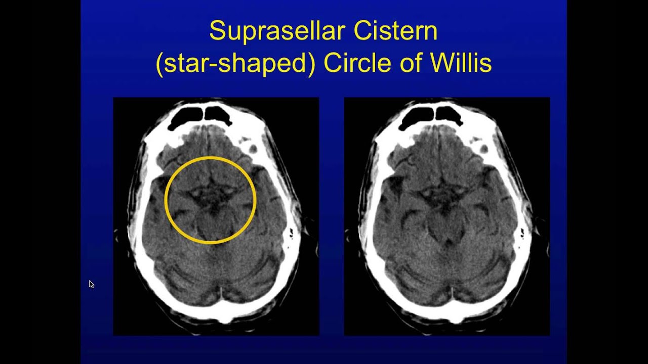
Head Ct Interpretation Made Easy Make It Simple Easy Circle Of Willis

Pin On Ct Scan In London

Closeup Of A Ct Scan With Brain Science And Education Mri Background Ad Affiliate Scan Brain Closeup Ct Ct Scan Science Education Photo Editing

Crash Ct Scan Guidance Radiology Imaging Ct Scan Brain Images

Pin On The 3rd Eye 6th Chakra Pineal Gland Seat Of The Soul

Stroke Ct Brain Scan Brain Scan Radiology Medical Imaging
Post a Comment for "ct scan brain"